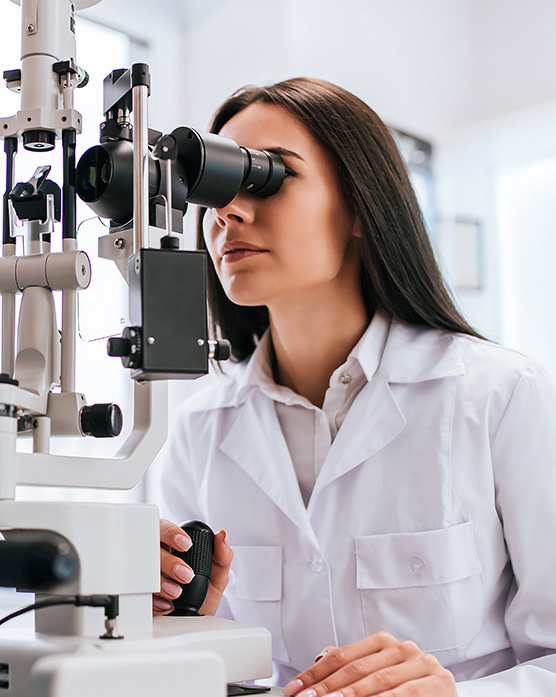
Our Diagnostic Devices
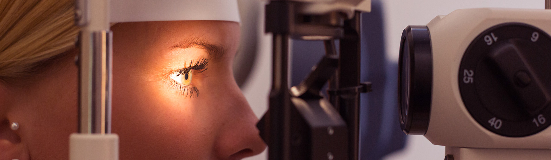
During the eye exam, we may use the following devices in our assessment of the back of the eye:
OCT (Optical Coherence Tomography)
OCT is a non-invasive diagnostic device used to create cross-sectional images of the retina and measure the integrity of the optic nerve. OCTs are often compared to ultrasounds, but instead of sound, they use light to generate their images.
The Zeiss Cirrus OCT offers highly regarded diagnostic abilities. In addition to assessing the retina and optic nerve, this OCT also measures and images the full anterior chamber (the front portion of the eye), allowing us to diagnose narrow angles, among other conditions.
Widefield fundus photography
Fundus photography can capture highly detailed images of the fundus (your eye’s interior surface). This non-invasive device is able to document diseases like diabetic retinopathy, diabetic macular edema, and age-related macular degeneration – and also provides an important baseline documentation of a healthy eye, so we can track changes occurring over time.
We are proud to use Zeiss Clarus Widefield Fundus Photography. Its images capture a significantly wider view than traditional fundus cameras while still maintaining the true colour of the fundus. It can also be aligned to avoid eyelids and eyelashes interfering in the view, resulting in clearer pictures and less time spent taking multiple photos.
Visual field
Peripheral vision (everything you see to the sides when looking straight ahead) is just as important as central vision (what you see in the direction your eyes are pointing).
We test peripheral vision on all patients over age 65 who are covered by OHIP. We may also check it in those patients whose peripheral vision is at higher risk. This could be due to glaucoma or other conditions affecting the optic nerve (which connects the eye to the part of the brain that processes vision); a history of stroke; or even the use of certain medications (e.g. hydroxychloroquine).
To assess peripheral vision, we use the Humphrey Visual Field Analyzer, the gold standard of visual field testing. The latest model, HFA3, gives us a significantly faster testing time. Combined with its trusted accuracy and precision, this makes the testing process easier and more comfortable than ever.